Anth 300 Forensic Anthropology
Spring 2008
Dr. Darlene Applegate
LAB 11: AGE ESTIMATION

INTRODUCTION
The analytical stage of forensic anthropology involves answering questions that lead to identification of the individual whose remains are being examined. The questions asked in developing a biological or demographic profile for an individual include the following:
OBJECTIVES
READINGS
TERMS
GENERAL INSTRUCTIONS
Carefully handle the instructional casts and bones laid out in the lab, being sure to keep the bones with their labels. Some of the casts are made of brittle plaster and will break if dropped. Some of the ends of the bones where you will be making measurements are very delicate and will crumble if handled improperly. Keep the materials on the bubble wrap to cushion them from the hard table surfaces, and wear gloves when working with real bones.
Record your responses to the questions in pencil on the answer sheet
provided
in the lab.
All estimates must include units (years, months) and be reported as a range (e.g., 14 to 16 years, less than 19 years, 3 years ± 9 months).
When working in a group, it is essential that all group members look at the bones and make the measurements. If you don't do this, you risk missing points on the lab and the lab test.
Use reference books in the lab as needed. Ask the instructor or assistant if you don't understand something.AGE ESTIMATION
Overview
The methods used to estimate age depend on the relative age of the individual. Developmental traits used to estimate the age of subadults include tooth eruption and epiphyseal union. In addition to fusion of the medial clavicle epiphysis and the cranial sutures, adult ages are estimated using degenerative traits like changes in the morphology of the pubic symphysis and auricular surface of the ilium, changes in the morphology of the sternal ends of the ribs, cranial suture morphology, dental attrition or wear of occlusional surfaces, bone resorption, osteon counting, and joint degeneration. Because developmental traits develop more regularly and consistently than degenerative traits, age estimates for subadults tend to be more accurate and within a smaller range of error than age estimates for adults and the elderly.
As with sex estimation, the more indicators used to determine age, the more accurate the results. However, a forensic anthropologist is analytically limited by the bones present and their condition. Age estimates are usually given as a range, such as 23-32 years, or with a range of error, such as 12 ± 2.5 years.
In this lab we will look at dental eruption, epiphyseal union, dental wear, and pubic symphysis morphology to estimate age. Again, what we are doing in lab for age estimation does not cover all skeletal indicators of age, but it will give you a good idea of how a forensic anthropologist estimates the age of an individual using the bones.
Dental Eruption
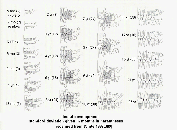
Eruption of deciduous (baby or milk) teeth and permanent
(adult) teeth occurs at fairly regular intervals during the
subadult
years of development (see the figure below, deciduous teeth are shaded;
this figure is also in your textbook and lab book). Therefore,
age estimation
of subadults based dental eruption is quite accurate. The image
belo, which is also in your textbook on page 223, will be used
to estimate age of skull casts
based on dental eruption.

Dental Attrition or Occlusional Wear
While tooth wear and permanent tooth loss can occur in subadults, these degenerative changes are usually associated with adults. Loss of permanent teeth and accompanying bone resorption of the alveolar bone of the maxilla and/or mandible are often associated with old age. Tooth wear or dental attrition most often occurs in adults, but the age of onset depends on diet and other environmental factors. This process leads to loss of outer white tooth enamel and exposure of the yellowish dentine of the pulp cavity, especially on the cusps of the teeth. The older an individual is, the more dentine is exposed due to tooth wear. The figure from White (1991) distributed in class illustrates this process for the maxillary and mandibular teeth. The exposed dentin is shown in black for the molars (left) to incisors (right) for one-fourth of the mouth.
Epiphyseal Union
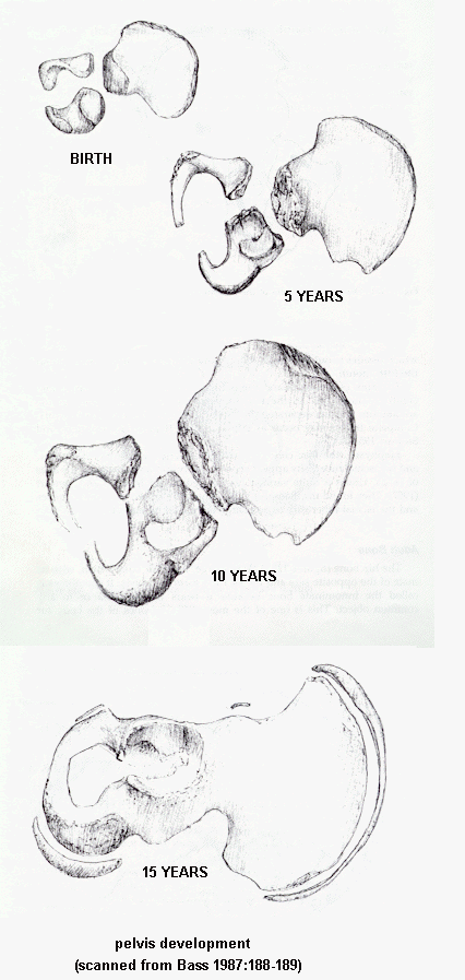
At birth a human has about 450 bones, over twice that of a human adult. A single bone in an adult, for example the os coxa, is usually a series of several bones in a subadult (see the figure below). During ontogenetic development the multiple bones fuse together into single bones, so that adults have 206 bones. This process is often referred to epiphyseal union.

The fusion of bone epiphyses to metaphyses occurs at
regular intervals during the course of ontogenetic development.
Because
of this regularity, epiphyseal union is a useful trait in aging
individuals,
especially subadults. While there is some sexual variation, with
female epiphysis fusion occurring earlier than in males, the ages of
epiphyseal
union are regular (see handout distributed in class).
The fusion of a particular bone is usually classified into stages: nonunion, one-quarter united, one-half united, three-fourths united, and fully fused or full union (White 1991:313).
We will examine epiphyseal union of the iliac crest of the os coxa
(based
on casts in lab), fusion of the os coxa bones (based on the figure
above),
the medial epiphysis of the clavicle (based on casts in lab), and the
long
bones (see France workbook page 220; subtract one year for females**).
Iliac Crest
The iliac crest of the coxa fuses in stages, with the corresponding
range of ages listed in the table below. Stage 1 represents
nonunion.
At Stage 2 the epiphysis is not fused and remains separate from the
metaphysis.
Stage 3 is partial union, and Stage 4 is complete union (Suchey, no
date).
| Cast | No. Pieces | Stage |
| Ii | 1 | 1 (nonunion) uneven, billowy metaphysis no epiphysis |
| Iii | 2 | 2 (nonunion with
separate
epiphysis) uneven, billowy metaphysis epiphysis present but not fused |
| Iiii | 2 | 3 (partial union at
anterior) uneven, billowy metaphysis ephiphyseal fusion anterior separate epiphysis posterior |
| Iiv | 1 | 3 (partial union) uneven metaphysis, less billowy partial epiphyseal fusion line visible |
| Iv | 1 | 3 (partial union) uneven metaphysis, less billowy partial epiphyseal fusion line visible |
| Ivi | 1 | 4 (complete union) no metaphysis fully fused epiphysis line visible |
| Ivii | 1 | 4 (complete union with
lipping from old age) no metaphysis fully fused epiphysis line visible arthritic lipping |
| Stage | Female | Male |
| 1 | 11 | 11-16 |
| 2 | 14-15 | 13-19 |
| 3 | 14-23 | 14-23 |
| 4 | 18-24 | 17-24 |
Medial Clavicle
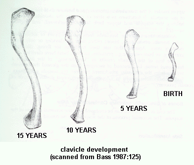
Not only does the clavicle increase in length during ontogenesis
(see
the figure below), but the epiphyses fuse at certain ages. The
medial
epiphysis of the clavicle is particularly useful in adult age
estimation
as it is one of the last epiphyses in the body to fuse.

The medial epiphysis of the clavicle fuses in stages, with the
corresponding
range of ages listed in the table below. Stage 1 represents
nonunion.
At Stage 2 the epiphysis is not fused and remains separate from the
metaphysis.
Stage 3 is partial union, and Stage 4 is complete union (Suchey, no
date).
| Cast | No. Pieces | Stage |
| Ci | 1 | 1 (nonunion) uneven, billowy metaphysis no epiphysis |
| Cii | 2 | 2 (nonunion with separate epiphysis) uneven, billowy metaphysis separate epiphysis |
| Ciii | 1 (1 is missing) | 3 (partial union at anterior) uneven, concave metaphysis separate epiphysis with partial fusion |
| Civ | 1 | 3 (partial union) uneven, concave metaphysis partial fusion of epiphysis small epiphysis |
| Cv | 1 | 3 (partial union) uneven, concave metaphysis partial fusion of epiphysis large epiphysis |
| Cvi | 1 | 4 (complete union) no metaphysis smooth epiphysis is fully fused slight line |
| Cvii | 1 | 4 (complete union with lipping from old age) no metaphysis smooth epiphysis is fully fused no line arthritic lipping |
| Stage | Female | Male |
| 1 | 11-23 | 11-25 |
| 2 | 16-21 | 16-22 |
| 3 | 16-33 | 17-30 |
| 4 | 20-34 | 21-31 |
Long Bone Epiphyseal Union
Using the handout provided in class, note the ages of
epiphyseal
union of the proximal and distal tibia and femur for white males. As
the figure illustrates, fusion of many other bones of the body can
be
used to determine age for subadults, but we will look at the tibia and
femur in this lab. For female specimens, subtract one year from the
reported age ranges.
Pubic Symphysis Degeneration
The surface morphology of the pubic symphysis changes with
age.
The pubic symphysis is regular, raised and "billowy" with an indistinct
margin in youth and irregular and depressed with a more distinct margin
in old age. The morphological transformation of the pubic
symphysis
follows a pattern that is divided up into phases. Suchey and
Brooks
identified 12 phases of pubic symphysis morphology for males and
females
and determined the average ages associated with each phase. We
will
use the figures in the textbook/workbook as well as
Suchey-Brooks plaster
casts of male and female pubis bones to estimate age (mean and standard
deviation) based on the pubic
symphysis.
Assignment
We will examine dental eruption, dental wear or attrition,
epiphyseal
union of the iliac, ilium, clavicle, tibia, and femur, and pubic
symphysis
morphology in this lab. Look at each specimen carefully and
record
your age estimates and explanations in pencil on the answer sheet. Be
sure each answer includes units (e.g., months, years).
DENTAL ERUPTION AND DENTAL ATTRITION
Examine the specimen labeled Dental A. Answer the specific questions regarding dental eruption and/or dental wear for this specimen. What is the age range for this individual?ILIAC CREST AND OS COXA FUSIONExamine the specimen labeled Dental B. Answer the specific questions regarding dental eruption and/or dental wear for this specimen. What is the age range for this individual?
Examine the specimen labeled Dental C. Answer the specific questions regarding dental eruption and/or dental wear for this specimen. What is the age range for this individual?
MEDIAL CLAVICLE FUSIONExamine the specimen labeled Os Coxa A and assume it is from a female individual. Using the Bass figure of os coxa fusion, what is the age range for this individual? At what stage of union is the anterior iliac crest (give roman numeral and arabic numeral)? What is the age range for this individual based on the iliac crest?
Examine the specimen labeled Os Coxa B and assume it is from a male individual. Using the Bass figure of os coxa fusion, what is the age range for this individual? At what stage of union is the anterior iliac crest (give roman numeral and arabic numeral)? What is the age range for this individual based on the iliac crest?
LONG BONE EPIPHYSEAL FUSIONExamine the specimen labeled Clavicle A, which is a complete clavicle. Assume this is a female specimen and no other clavicle bone parts were present. At what stage of fusion is the medial clavicle (give roman numeral and arabic numeral)? What is the age estimate for this individual?
Examine the specimen labeled Long Bone A and assume it is from a female individual. What is the age estimate for this individual?PUBIC SYMPHYSIS DEGENERATIONExamine the specimen labeled Long Bone B and assume it is from a male individual. What is the age estimate for this individual?
Examine the specimen labeled Pubis A. Are these specimens from a male or female? Using the Suchey-Brooks casts for the appropriate sex, what is the phase of pubic symphysis morphology? What is the age estimate (expressed as the mean and standard deviation) for this individual?Examine the specimen labeled Pubis B. Are these specimens from a male or female? Using the Suchey-Brooks casts for the appropriate sex, what is the phase of pubic symphysis morphology? What is the age estimate (expressed as the mean and standard deviation) for this individual?
REFERENCES
Bass, W. M. (1987) Human Osteology: A Laboratory and Field Manual
(3rd ed.). Missouri Archaeological Society, Columbia.
Byers, Stephen N.
2005 Introduction to Forensic
Anthropology: A Textbook. Allyn and Bacon, Boston.
Byers, Stephen N. and Susan M.T. Myster
2005 Forensic Anthropology
Laboratory Manual. Allyn and Bacon, Boston.
France, D. L. (2003) Lab Manual and Workbook for Physical Anthropology (5th ed.). West / Wadsworth, Belmont, CA.
White, T. D. (1991) Human Osteology. Academic Press, San
Diego.