
INTRODUCTION
The analytical stage of forensic anthropology involves answering questions that lead to identification of the individual whose remains are being examined. The questions asked in developing a biological or demographic profile for an individual include the following:
OBJECTIVES
READINGS
TERMS
Taphonomy is the study of the fossilization process or the
biological or geological processes that
affect
the condition, preservation or location of skeletal remains after an
individual dies. By
definition,
taphonomic processes are postmortem,
meaning they affect the
hard
tissues after
death. The effects of taphonomic processes on bones
must be distinguished from skeletal attributes used in developing a
biological
profile.
BIOLOGICAL PROCESSES
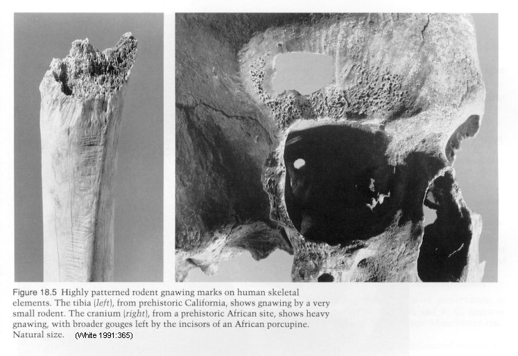
Biological taphonomic processes are related to plant and animal activity and include rodent gnawing, carnivore gnawing, carnivore digestion, algal growth and root etching.
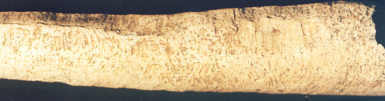
Rodent gnawing leaves paired, U-shaped parallel incisions on
bones. Rodents typically gnaw on the projections of bones
(crests,
tuberosities, condyles, processes, etc.), but the incisions also occur
on low-relief surfaces of bones. The fact that rodent gnawing
marks
are paired distinguishes them from carnivore gnawing, and the fact that
rodent gnawing marks follow the contour of a bone distinguishes them
from
cut-marks or incised sharp-force traumas.
When rodents gnaw in opposite directions on a bone surface, they may
create what appears to be a ridge or crest, as pictured in the upper
left
of the bone below.
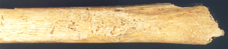
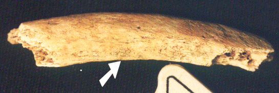
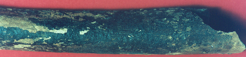
Carnivore gnawing leaves single, U-shaped incisions or
"channels"
on bones that should not be confused with incised sharp-force
traumas.
Carnivores also leave small round depressed fractures where their
canine
teeth puncture the cortical bone. In addition, they gnaw the ends
of bones and bone projections, exposing spongy bone; carnivores crush
bone
shafts in order to extract marrow, creating "spiral" fractures.
Note
the punctures on the bone below.
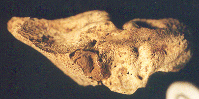
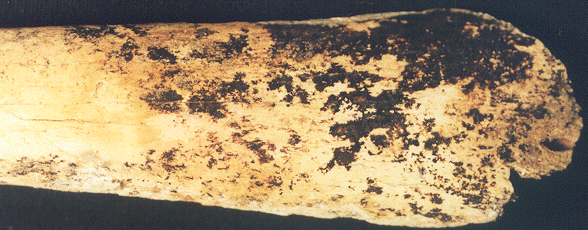
Carnivore digestion results in pitting and dissolution of
bone,
which often exposes spongy bone. A bone that is pitted by
carnivore
digestion is shown below. This taphonomic alteration should not
be
confused with periosteal reactions.
Algal growth is evidenced by the accumulation of
discontinuous
green spots or continuous green areas on bone surfaces. Algae
grows
on bones in moist, warm environments. It should not be confused
with
copper staining from buttons or other evidence. The arrow on the
rib fragment below points to green algal growth.
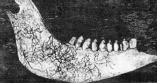
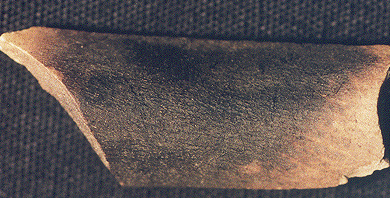
The weak acids released by plants through their roots can leave root
etching marks on bone surfaces. The etching is typically into
the
cortical bone and forms a dendritic pattern. The etching actually
creates subsurface linear patterns on the bone surfaces, though in the
photo below of an etched sheep mandible it appears that the alteration
is deposited on top of the bone surface.
http://hoopermuseum.earthsci.carleton.ca/taphonomy/BONMOD4.HTM
GEOLOGICAL PROCESSES
Geological taphonomic processes relate to erosional and weathering agents and include element/mineral deposition, water erosion, wet-dry or freeze-thaw cycling, sun exposure, and burning.
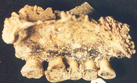
Elements and minerals may be deposited on bone surfaces as a result of precipitation of those materials from groundwater contact. Common deposits include manganese, calcite or calcium carbonate, and iron or iron oxides.
Manganese deposition is characterized by discontinuous,
amorphous
patches of dull black material on bone surfaces. It should not be
confused by carbonization from burning, which is usually more glossy or
reflective.
Calcite (calcium carbonate) deposition is characterized by
discontinuous or
continuous,
amorphous patches of grayish, brittle, flaky material on bone
surfaces.
It should not be confused with arthritic osteophyte development or
dental
calculus.
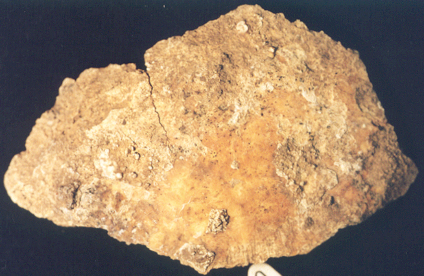
Iron and iron oxide deposition is characterized by
discontinuous,
amorphous, rust-colored or blackened areas or spots on bone
surfaces.
It should not be confused with carbonization from burning.
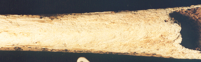
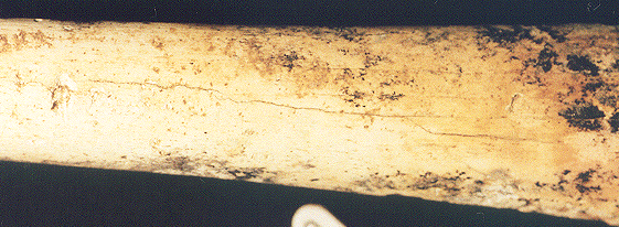
Water erosion results in rounding and smoothing of bone edges
and projections as well as cortical bone loss.
Repeated cycles of wet-dry or freeze-thaw conditions
cause
exfoliation or delamination, or the loss of cortical bone as
well
as the development of longitudinal fractures, which are breaks
oriented
parallel to bone shafts or surfaces. The images below represent
exfoliation
and longitudinal fracturing.
Exposure to the sun causes bones to become bleached
or
whitened, such as the cow skull below.
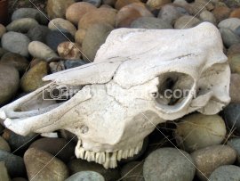
http://www.istockphoto.com/file_thumbview_approve/187260/2/Where_s_the_Beef_.jpg
Natural fires can alter bones in the same ways as vehicular or
residential
fires. Bones initially become fractured and blackened or carbonized
(as shown below on animal bone). As the burning progresses,
bones
become increasingly fragmented, fractured and whitened or calcined.
ASSIGNMENT
Using the above descriptions and comparative materials in the lab,
examine
each of the real bone specimens. For each specimen, identify the
skeletal element and side (for paired bones). Identify the
taphonomic
agent(s) that affected the bones and, if asked, describe the effects
(e.g.,
type of bone alteration, location of alteration, extent of alteration)
of each agent.
REFERENCE
White, T. D. (1991) Human Osteology. Academic Press, San
Diego.
Unless otherwise noted, all photographs are by Darlene Applegate.