
INTRODUCTION
First, recall the goals of the archaeological excavations at Hilltopper Shelter. One research question concerns when the site was occupied. Is Hilltopper Shelter a single component historic period site or a multicomponent site that also represents prehistoric occupations? Can a specific range of years be specified for each occupation? The other research question relates to the function of the site during each temporally-distinct occupation. How was the site used during each occupation?
In the previous labs we considered how lithic,
ceramic/pottery,
archaeobotanical, and zooarchaeological artifacts might be used to
answer
these questions. In this lab, we examine how human remains can be
used to address these questions. Our osteological analysis will proceed
with these questions in mind. How can human remains be used to
determine
time and period of occupation? How can human remains be used to
determine
site function? Osteological/bioarchaeological analysis is
considered
in this lab.
OSTEOLOGICAL/BIOARCHAEOLOGICAL ANALYSIS
Osteology is the study of the human skeleton, and
bioarchaeology is the study of human remains (e.g., skeletal remains,
paleofeces,
soft tissues) from archaeological contexts. In this lab, we
consider
a specific type of human remains, skeletal remains, recovered from
archaeological
contexts at Hilltopper Shelter. Bioarchaeologists who study human
skeletal remains typically are concerned with determining race, sex,
age,
and stature; estimating the minimum number of individuals; and
assessing
trauma, pathology, anomalies, and taphonomic alterations.
RACE DETERMINATION
Patterns of geographic variation of the human skeleton are used to identify the race or ancestry of an individual. Most bioarchaeologists use a three-race model that includes Mongoloid, Negroid, and Caucasoid races. Native Americans are typically included in the Mongoloid race.
Skeletal indicators of race focus primarily on skull and dental traits. Racial indicators on the skull are both nonmetric and metric traits and include robusticity, lengths and widths of skull features, shapes of skull features, and unique population-specific dental features.
The following table summarizes typical
expressions
of 28 metric and nonmetric skull traits for the three human
races.
A guide to assessing these traits follows the table.
| TRAIT | CAUCASOID | NEGROID | MONGOLOID |
| 1. cranial index | 75 to 80, mesocranic | less than 75, dolicocranic | greater than 80, brachycranic |
| 2. sagittal contour | arched | flat with bregmatic or post-bregmatic depression | arched |
| 3. keeling of skull vault | absent | absent | present |
| 4. total facial index | greater than 90, narrow to very narrow | less than 85, broad to very broad | 85 to 90, medium or average |
| 5. facial profile | orthognathic (straight, flat) | prognathic (projecting), especially in the alveolar area | intermediate to mostly orthognathic |
| 6. nuchal ridge profile | pinched and prominent | slightly pinched | rounded |
| 7. base chord | long | long | short |
| 8. suture pattern | simple | simple | complex |
| 9. metopic suture | present | absent | absent |
| 10. wormian bones | absent | absent | present |
| 11. eye orbit shape | angular and sloping | square or rectangle | rounded and non-sloping |
| 12. lower eye border | receding | receding | projecting |
| 13. nasal index | less than 48, leptorrhinic (narrow) | greater than 53, platyrrhinic (wide) | 48 to 53, mesorrhinic (intermediate) |
| 14. nasal cavity shape | tear shaped | rounded and wide | oval shaped |
| 15. nasal bones | "tower shaped," narrow and parallel from anterior, slightly arched in profile | "Quonset hut shaped," wide and expanding from anterior, no arch in profile | "tented," narrow and expanding from anterior, arched in profile |
| 16. nasal overgrowth | absent | absent | present |
| 17. nasal sill or dam | present | absent | absent |
| 18. lower nasal spine | large and sharp | small | small |
| 19. zygomatic arches | narrow and retreating | medium to large and retreating | projecting |
| 20. external auditory meati | round | round | oval |
| 21. palate shape | triangular | rectangular | parabolic or horseshoe shaped |
| 22. palate suture | irregular | irregular | straight |
| 23. occlusion | slight overbite | slight overbite | edge-to-edge or even |
| 24. central incisors | blade shaped | blade shaped | shovel shaped |
| 25. ascending ramus of mandible (shape) |
pinched at midsection | back slanted | wide and vertical |
| 26. ascending ramus of mandible
(projection) |
non-projecting |
projecting |
non-projecting |
| 27. gonial angle | slightly flared | not flared | slightly flared |
| 28. chin profile | prominent and projecting | rounded | slightly projecting |
1. CRANIAL INDEX: Use the spreading caliper. Measure the maximum breadth of the skull from euryon (eu) to euyron (eu). Measure the length of the skull from glabella (g) to opisthocranion (op). Divide the cranial breadth by the cranial length and multiply by 100. (See figures below for landmarks.)
2. SAGITTAL CONTOUR: Holding the skull in profile, examine the contour of the cranium along the sagittal suture.
3. KEELING OF SKULL VAULT: Holding the skull in anterior position, examine the contour of the cranium. Keeling is a pinched appearance along the sagittal suture.
4. TOTAL FACIAL INDEX: Use the sliding caliper to measure the maximum heighth of the face from nasion (n) to gnathion (gn). Use the spreading caliper to measure the maximum width of the face from zygion to zygion (zy). Divide the facial height by the facial width and multiply by 100. (See figures below for landmarks.)
5. FACIAL PROFILE: Holding the skull in profile, gently "place one end of your pencil on or near the anterior nasal spine (on the midline of the skull) at the base of the nasal aperture [nasal cavity]. Lower the pencil toward the face so that the pencil will touch the chin" (Bass 1987:87). If the pencil hits the alveolar area of the mouth, the face is prognathic. If the pencil extends to the chin, the face is orthognathic. "Caucasoids have a 'flat' (orthognathous) face in the dental area along the midline. This is the opposite of the Negroid face, which exhibits protrusion of the mouth region, known as prognathism. ... Negroids are noted for alveolar prognathism, or an anterior protrusion, of the mouth region. A pencil or ballpoint pen placed with one end on the nasal spine (midline at base of nasal aperture) will not touch the chin (the teeth protrude too far forward)" (Bass 1986:87).
6. NUCHAL RIDGE PROFILE: Holding the skull in profile, examine the nuchal ridge and note the shape.
7. BASE CHORD: Holding the skull in inferior view, examine the distance between opisthion and opisthocranion.
8. SUTURE PATTERN: Examine the pattern of the cranial sutures (sagittal, cornonal, squamosal, lambdoidal) and describe the pattern as simple (not very convoluted) or complex (very convoluted).
9. METOPIC SUTURE: Examine the frontal bone superior to the nasal bones for evidence of a short suture known as the metopic suture.
10. WORMIAN BONES: Examine the lambdoidal suture and look for small bones within the suture line. These bones are called wormian bones.
11. EYE ORBIT SHAPE: Examine the eye orbits from the anterior view. Describe the overall shape as rounded or squared. If the eye orbits are rounded, examine the top border to see if it is level or if it slopes laterally.
12. LOWER EYE BORDER. Examine the skull in profile, gently placing a pencil vertically across the eye orbit. If the pencil is a vertical plane, then the lower eye border is projecting. If the pencil is not a vertical plane, then the lower eye border is not projecting.
13. NASAL INDEX: Using the sliding caliper, measure the maximum breadth of the nasal cavity (at right angles to the nasal height), from alare to alare (al). Measure the nasal height from nasion (n) to nasospinale (ns). Divide the nasal breadth by the nasal height and multiply by 100. (See figures below for landmarks and measurement information.)
14. NASAL CAVITY SHAPE: Examine the overall shape of the nasal cavity from the anterior view.
15. NASAL BONES: Examine the shape of the nasal bones from the anterior and lateral views. From the anterior view, check the width of the bones and whether or not they expand outward from superior to inferior. For the lateral view, check if the bones arch downward (concave up).
16. NASAL OVERGROWTH: Examine the nasal bones in lateral view. An overgrowth is present if the inferior ends of the nasal bones overhang the superior edge of the nasal cavity.
17. NASAL SILL OR NASAL DAM: "Carefully observe the base of the nasal aperture [nasal cavity or opening]. With your pencil or ballpoint pen resting against the bone of the maxilla just below the nasal opening, try to run the pencil or pen gently into the nasal opening. In Caucasoids there is usually a dam (nasal sill) that will stop the pen or pencil. In Negroid skulls there is no dam or nasal sill, and the pen easily will glide into the nasal aperture. Mongoloid skulls will range between these two extremes" (Bass 1986:83). Be extremely careful when inserting a pen or pencil into the nasal cavity to avoid bone damage.
18. LOWER NASAL SPINE: Holding the skull in lateral view, examine the lower nasal spine that extends from the inferior edge of the nasal cavity. Describe the shape.
19. ZYGOMATIC ARCHES: "Hold the skull with the occipital region in your hand and the facial area up. Place a pencil across the nasal aperture [nasal cavity]. Now try to insert your index finger between the cheek (zygomatic) bones and the pencil. Caucasoids have a face that comes to a point along the midline and cheek bones that do not extend forward. This will allow you to insert your finger between the cheek bones and the pencil without knocking the pencil off. Mongoloids have a much flatter face (the cheek bones extending much further forward), and it is difficult to insert your finger between the pencil and the cheek bones on a Mongoloid skull without knocking the pencil off" (Bass 1986:83).
20. EXTERNAL AUDITORY MEATI: Holding the skull in lateral views, examine the overall shapes of the external auditory meati.
21. PALATE SHAPE: Holding the skull in inferior view, examine the palate area, which includes the maxillae and palatines. Describe the overall shape.
22. PALATE SUTURE: Holding the skull in inferior view, examine the middle portion of the suture between the maxillae and palatines. Describe the shape.
23. OCCLUSION: Holding the skull in lateral view, examine the occlusion of the upper and lower incisors. If the maxillary incisors are anterior relative to the mandibular incisors, this is an overbite. If the maxillary and mandibular incisors meet evenly, this is edge-to-edge occlusion.
24. CENTRAL INCISORS: Holding the mandible in superior view and/or the maxillae in inferior view, examine the shape of the central incisors. Shovel-shaped incisors have posterior-oriented projections.
25. ASCENDING RAMUS OF MANDIBLE (SHAPE): Holding the
mandible in
lateral view, examine the overall shape of the ascending ramus.
26. ASCENDING RAMUS OF MANDIBLE (PROJECTION): Holding
the mandible in posterior view, examine the posterior edge of the
ascending ramus and look for a bony projection toward the midline.
27. GONIAL ANGLE: Holding the mandible in anterior view, examine the gonial angle to see if it is rounded or outward flaring.
28. CHIN: Holding the mandible in lateral view, examine
the relative projection of the chin.
SEX DETERMINATION
The sex of an individual is determined, when soft tissue is not present, by a number of skeletal indicators including the pelvic girdle, skull, and long bones. Of course, the more indicators used to determine sex, the more accurate the results. However, a bioarchaeologist is analytically limited by the bones present and the condition of the bones. Sex determination is very difficult for subadults.
The pelvic girdle is often cited as the best
skeletal
element with which to determine sex. We are using the pubis bone,
various os coxae features, and the overall shape of the pelvic girdle
to
estimate sex.
|
|
|
|
| ventral arc of pubis | present | absent |
| pubis body width | > 40 mm | < 25-30 mm |
| subpubic angle | > 90 degrees | < 90 degrees |
| obturator foramen | small and triangular | large and ovoid |
| greater sciatic notch | wide, >90 degrees | narrow, deep, <90 degrees |
| auricular surface | high relief, with pre-auricular sulcus | low relief , lacking pre-auricular sulcus |
| acetabulum | small | large |
| pelvic inlet | broad, circular | narrow, heart-shaped |
After the pelvis, the skull is the bone commonly
used to determine sex. We are examining a number of skull
features
that are indicators of sex. Many of these features are relative,
meaning that the male:female differences are most easily observed when
looking at one skull relative to another. Skull features that are
used to distinguish males and females are listed below.
| Trait | Female | Male |
| supraorbital ridge/torus | less prominent | more prominent |
| upper edge eye orbit | sharp | blunt |
| eye orbit shape | rounded | square |
| palate | smaller | larger |
| teeth | smaller | larger |
| chin | rounded with midline point, V-shaped | square, U-shaped |
| gonial angle | non-projecting | projecting or flaring |
| cranial vault | smaller, smoother | larger, rougher |
| frontal bossing* | present | absent |
| parietal bossing* | present | absent |
| muscle ridges (nuchal) | gracile | robust |
| zygomatic process | not expressed beyond zygomatic arch | expressed beyond zygomatic arch and beyond external auditory meatus as crest |
| mastoid process | smaller, short | larger, broad |
| occipital condyles | smaller | larger |
Ordinarily, the limb bones alone are not used for sexing unless absolutely necessary. But characteristics of the limb bones can be used (alone or in conjunction with examination of the pelvis and skull) for sex estimation.
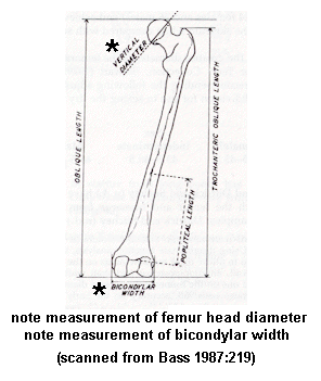
Two metric traits of the femur used to estimate sex are maximum diameter of the head (vertical diameter) and bicondylar width. The figure below shows how to measure these traits.

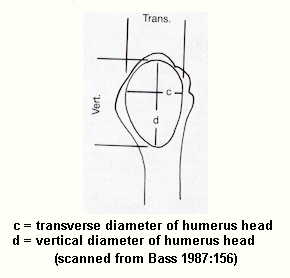
Three metric traits of the humerus used to estimate sex are maximum diameter of the head (transverse and vertical), epicondylar width (maximum width of the distal end), and maximum length. The figure below shows how to measure two of head diameters.

These metric traits vary between males and
females
in the following ways. All measurements are in mm. If the
race
you need is not present in the table, use another race.
| Trait | Female | Probably Female | Indeterminate | Probably Male | Male |
| femur head maximum diameter (Negroid) | <41.5 | 41.5-43.5 | 43.5-44.5 | 44.5-45.5 | >45.5 |
| femur head maximum diameter (Caucasoid) | <42.5 | 42.5-43.5 | 43.5-46.5 | 46.5-47.5 | >47.5 |
| femur bicondylar width (Negroid) | <72 | 72-74 | 74-76 | 76-78 | >78 |
| humerus head max diameter vertical (race?) | <43 | - | 44-46 | - | >47 |
| Trait | Female | Male |
| femur head maximum diameter (Caucasoid) | 43.8 | 49.7 |
| femur head maximum diameter (Negroid) | 41.5 | 47.2 |
| humerus head maximum diameter vertical (race?) | 42.7 | 48.8 |
| humerus head maximum diameter transverse (race?) | 37.0 | 44.7 |
| humerus head maximum diameter transverse (Native American) | 38-39 | 43-44 |
| humerus maximum length (Negroid) | 305.9 | 339.0 |
| humerus epicondylar width (Negroid) | 56.8 | 63.9 |
AGE ESTIMATION
The methods used to estimate age depend on the relative age of the individual. Developmental traits used to estimate the age of subadults include tooth eruption and epiphyseal union. In addition to fusion of the medial clavicle epiphysis and the cranial sutures, adult ages are estimated using degenerative traits like changes in the morphology of the pubic symphysis and auricular surface of the ilium, changes in the morphology of the sternal ends of the ribs, cranial suture morphology, dental attrition or wear of occlusional surfaces, bone resorption, osteon counting, and joint degeneration. Because developmental traits develop more regularly and consistently than degenerative traits, age estimates for subadults tend to be more accurate and within a smaller range of error than age estimates for adults and the elderly.
As with sex estimation, the more indicators used to determine age, the more accurate the results. However, a bioarchaeologist is analytically limited by the bones present and their condition. Age estimates are usually given as a range, such as 23-32 years, or with a range of error, such as 12 ± 2.5 years.
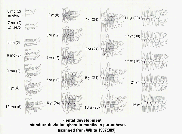
Eruption of deciduous (baby or milk) teeth and permanent (adult) teeth occurs at fairly regular intervals during the subadult years of development (see the figure below; deciduous teeth are shaded). Therefore, age estimation of subadults based dental eruption is quite accurate.

At birth a human has about 450 bones, over twice that of a human adult. A single bone in an adult is usually a series of several bones in a subadult. During ontogenetic development the multiple bones fuse together into single bones, so that adults have 206 bones. This process is often referred to epiphyseal union. The fusion of bone epiphyses to metaphyses occurs at regular intervals during the course of ontogenetic development. Because of this regularity, epiphyseal union is a useful trait in aging individuals, especially subadults. While there is some sexual variation, with female epiphysis fusion occurring earlier than in males, the ages of epiphyseal union are regular.
While tooth wear and permanent tooth loss can occur in subadults, these degenerative changes are usually associated with adults. Loss of permanent teeth and accompanying bone resorption of the alveolar bone of the maxilla and/or mandible are often associated with old age. Tooth wear or dental attrition most often occurs in adults, but the age of onset depends on diet and other environmental factors. This process leads to loss of outer white tooth enamel and exposure of the yellowish dentine of the pulp cavity, especially on the cusps of the teeth. The older an individual is, the more dentine is exposed due to tooth wear.
The surface morphology of the pubic symphysis
changes
with age. The pubic symphysis is regular, raised and "billowy"
with
an indistinct margin in youth and irregular and depressed with a more
distinct
margin in old age. The morphological transformation of the pubic
symphysis follows a pattern that is divided up into phases.
Suchey
and Brooks identified 12 phases of pubic symphysis morphology for males
and females and determined the average ages associated with each phase.
STATURE ESTIMATION
The stature or height of an individual is useful information for describing past human populations. Before estimating stature, one must determine the race, sex, and age of the individual as stature varies with the variables. Stature estimates are just that, estimates. They are not exact and should always be expressed with a range of error. Stature estimates are usually calculated in centimeters.
The lengths of individual long bones are used to
estimate stature. Bone length and stature tables for a number of
human populations have been published. Below are published
regression
formulas for stature estimation (Bass 1986:156-157, 163-164, 221-222,
238,
244).
| BONE | RACE | MALE EQUATION | FEMALE EQUATION |
| Femur | Caucasoid | 2.32 * femur + 65.53 ± 3.94 cm | 2.47 * femur + 54.10 ± 3.72 cm |
| Femur | Negroid | 2.10 * femur + 72.22 ± 3.91 cm | 2.28 * femur + 59.76 ± 3.41 cm |
| Femur | Mongoloid | 2.15 * femur + 72.57 ± 3.80 cm | not available |
| Tibia | Caucasoid | 2.42 * tibia + 81.93 ± 4.00 cm | 2.90 * tibia + 61.53 ± 3.66 cm |
| Tibia | Negroid | 2.19 * tibia + 85.36 ± 3.96 cm | 2.45 * tibia + 72.56 ± 3.70 cm |
| Tibia | Mongoloid | 2.39 * tibia + 81.45 ± 3.24 cm | not available |
| Fibula | Caucasoid | 2.60 * fibula + 75.50 ± 3.86 cm | 2.93 * fibula + 59.61 ± 3.57 cm |
| Fibula | Negroid | 2.34 * fibula + 80.07 ± 4.02 cm | 2.49 * fibula + 70.90 ± 3.80 cm |
| Fibula | Mongoloid | 2.40 * fibula + 80.56 ± 3.24 cm | not available |
| Humerus | Caucasoid | 2.89 * humerus + 78.10 ± 4.57 cm | 3.36 * humerus + 57.97 ± 4.45 cm |
| Humerus | Negroid | 2.88 * humerus + 75.48 ± 4.23 cm | 3.08 * humerus + 64.67 ± 4.25 cm |
| Humerus | Mongoloid | 2.68 * humerus + 83.19 ± 4.16 cm | not available |
| Ulna | Caucasoid | 3.76 * ulna + 75.55 ± 4.72 cm | 4.27 * ulna + 57.76 ± 4.30 cm |
| Ulna | Negroid | 3.20 * ulna + 82.77 ± 4.74 cm | 3.31 * ulna + 75.38 ± 4.83 cm |
| Ulna | Mongoloid | 3.48 * ulna + 77.45 ± 4.66 cm | not available |
| Radius | Caucasoid | 3.79 * radius + 79.42 ± 4.66 cm | 4.74 * radius + 54.93 ± 4.24 cm |
| Radius | Negroid | 3.32 * radius + 85.43 ± 4.57 cm | 3.67 * radius + 71.79 ± 4.59 cm |
| Radius | Mongoloid | 3.54 * radius + 82.00 ± 4.60 cm | not available |
MINIMUM NUMBER OF INDIVIDUALS
The miniumum number of individuals in an
archaeological
context is determined by totaling the number of unique skeletal
elements
(e.g., left humerus, right parietal, left patella, right maxillar third
molar) in each age-sex (e.g., adult male, adult female, subadult male,
subadult female) group.
OTHER ANALYSES
Bioarchaeologists also are interested in trauma,
pathology, anomalies, and taphonomic alterations. However, these
analyses will not be covered in our lab assignment.
OCCUPATION HISTORY
None of the human remains was submitted for
radiocarbon dating. Therefore, you do not need to consider
osteological
evidence related to the question of when the site was occupied.
SITE FUNCTION
Several issues related to site function may be addressed by osteological analysis of skeletal remains recovered from the shelter.
First, what activities may have occurred at the site? If human remains are recovered from grave features or from midden deposits, then burial or interrment activities occurred at the site. If special tools, features, or structures (e.g., chert knives and scrapers, crematory basins, charnal houses) associated with preparing the body for burial are associated with the remains, then mortuary treatment activities occurred at the site.
Second, what type of group used the site? Human remains may be used to identify the race of people who used the site. Human remains may be used to characterize the demographic makeup (e.g., sex distribution, age distribution) of the group(s) who used the site.
ASSIGNMENT: Using
reference materials and comparative collections in the lab, identify
the
skeletal element represented by each specimen. Record the
specimen
number, provenience, bone element (if possible), sex, age, and any
other
attributes you deem relevant to answering the research questions.
SITE REPORT
The results of the osteological analysis must be described in the site report. The osteological analysis section usually includes descriptive text, data tables, data figures, and quantitative analysis. I suggest a paragraph (with supporting figures and tables, as appropriate) describing the composition, distribution and preservation of the human remains from the site.
A brief paragraph indicating that the human remains were not used to estimate periods of site occupation should follow.
Follow this with paragraphs (plus supporting figures and tables) on the nature of site use during each occupation. What types of activities, based on human remains, occurred at the site? What types of groups used the site during each occupation?
ASSIGNMENT:
Compose
the osteological analysis section of the final site report.
Follow
the stylistic format of the existing portions of the site report.
Click here for more details.